 ALERT
ALERT **Important for iLab Users**
The Help link in iLab opens an email to contact Support. To submit or track requests, visit: Submit and Track Support Requests
The Microscopy and Histology Core offers comprehensive support for imaging experiments involving tissue explants, fixed tissues, and live/fixed cells. We provide a range of services, including equipment training, consultation, and assistance with all aspects of the imaging workflow, from sample preparation to microscopy image acquisition and image analysis.
Our core facility is equipped with state-of-the-art microscopy systems and histology equipment to ensure high-quality sample preparation & imaging results. Whether you are conducting basic research, studying disease models, or performing advanced imaging experiments, our experienced team is here to help.
The primary objective of this Core Facility is to cater to the research needs of the Health Science Center (HSC) and the broader scientific community in Fort Worth. We provide cutting-edge facilities that are open to faculty members, staff, and graduate students, enabling them to carry out their research effectively.
Moreover, our commitment extends beyond our institution as we offer our facilities and expertise to external individuals and organizations. By doing so, we aim to contribute to the advancement of the wider research community and promote collaboration among various institutions.
MICROSCOPY INSTRUMENTS
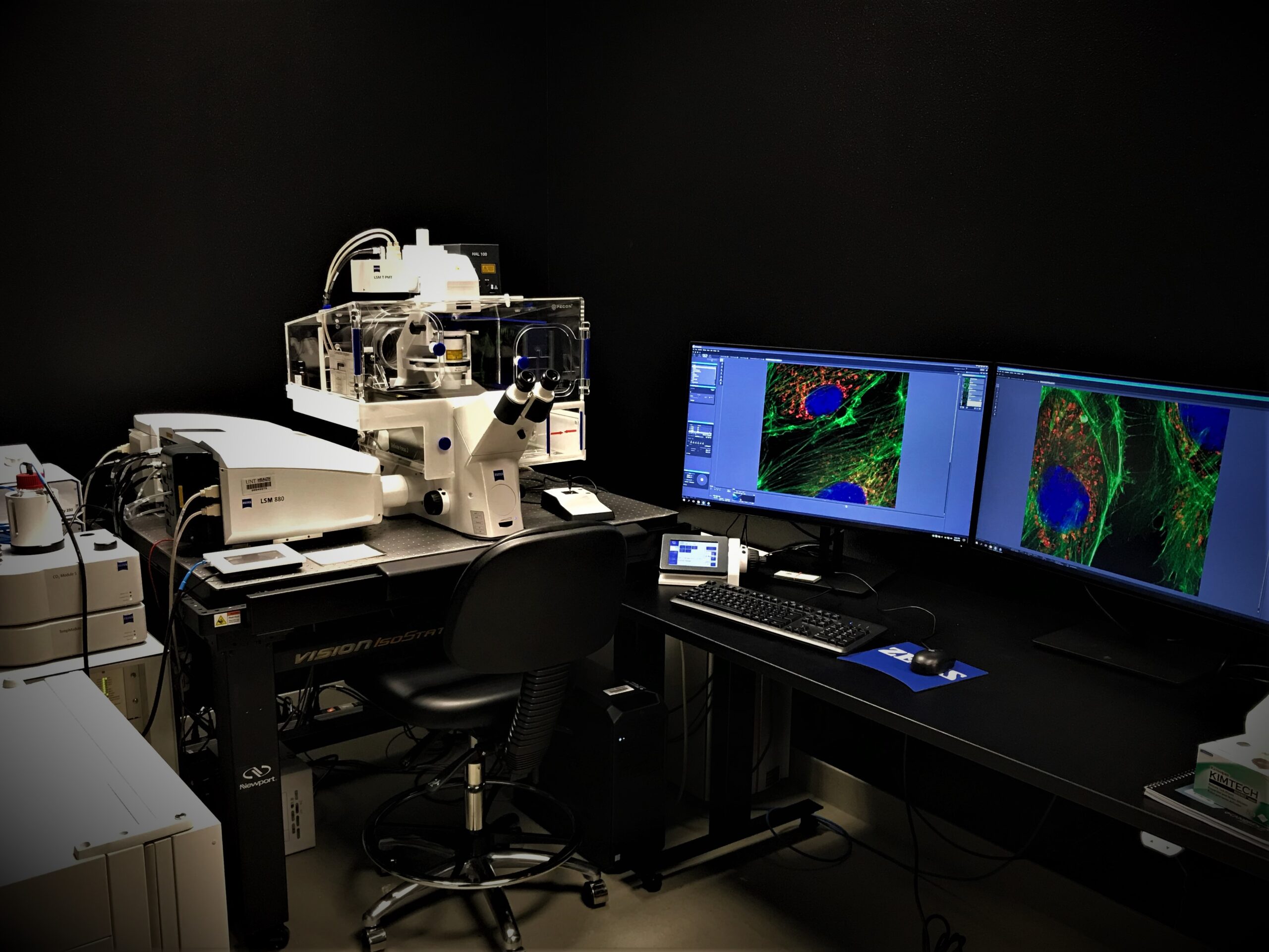
Zeiss LSM 880 Confocal (inverted) with AiryScan Super Resolution:
This inverted confocal laser scanning microscope is equipped with six laser lines, a variety of objectives, filter sets and detection systems that offers simultaneous 5-channel, super-resolution, ultrafast imaging of fixed tissues and live-cell.
Zeiss LSM 880 Confocal (upright) with 2-Photon excitation laser:
This upright confocal laser scanning microscope is equipped with six laser lines, a tunamble 2-Photon excitaion laser, variety of objectives, filter sets and detection systems that offers simultaneous 5-channel confocal imaging and intravital imaging.
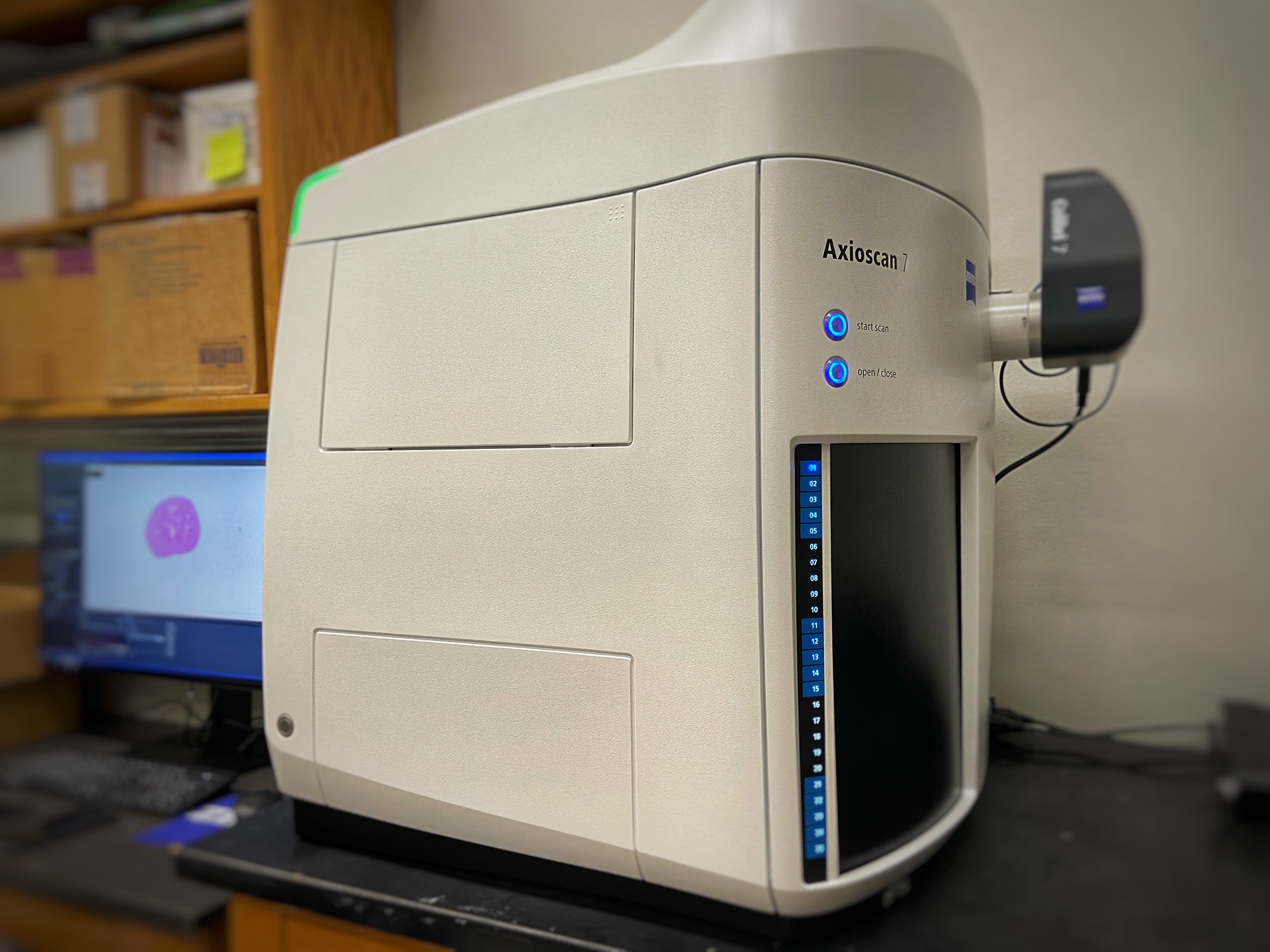
Zeiss Axioscan 7: This brand-new instrument in the core allows to digitize specimens in a reliable, reproducible way to create high-quality virtual microscope slides. With the availability of the highest quality objectives and swift and reproducible 7 color LED illumination with corresponding filter cube, as well as other advanced components like accurate autofocus and automatic sample detection, digitization of microscopy specimens is truly reimagined.
Keyence BZ-X800 All-in-One Fluorescence Microscope:
The Keyence BZ-X800 consists of fully motorized control system fitted withvadvanced image processing and analysis functions, five filter cubesvand a selection of different high-quality objectives with live-cell imaging capability.
Image Analysis Workstation: The workstation is a high-performance processing system for users with Zeiss Zen Black/Blue, Huygens, FIJI/ImageJ, Napari and Cell profiler image processing and analysis software.
Huygens Software: This image processing and analysis software uses advanced algorithm for deconvolution which significantly augment the resolution of the microscopy images. It has features like object analyzer, surface renderer, movie maker to name a few.
HISTOLOGY Instruments and Services:
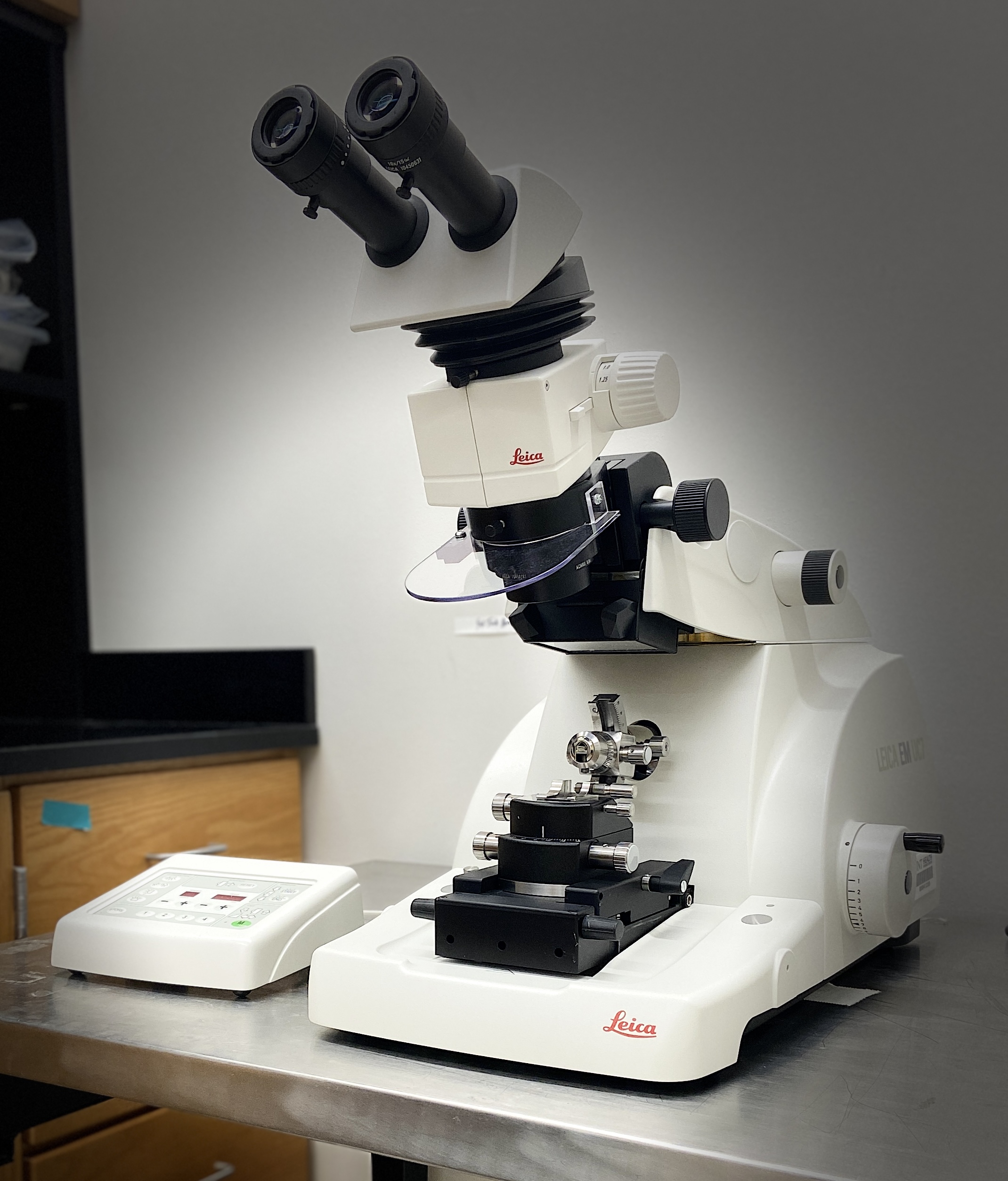
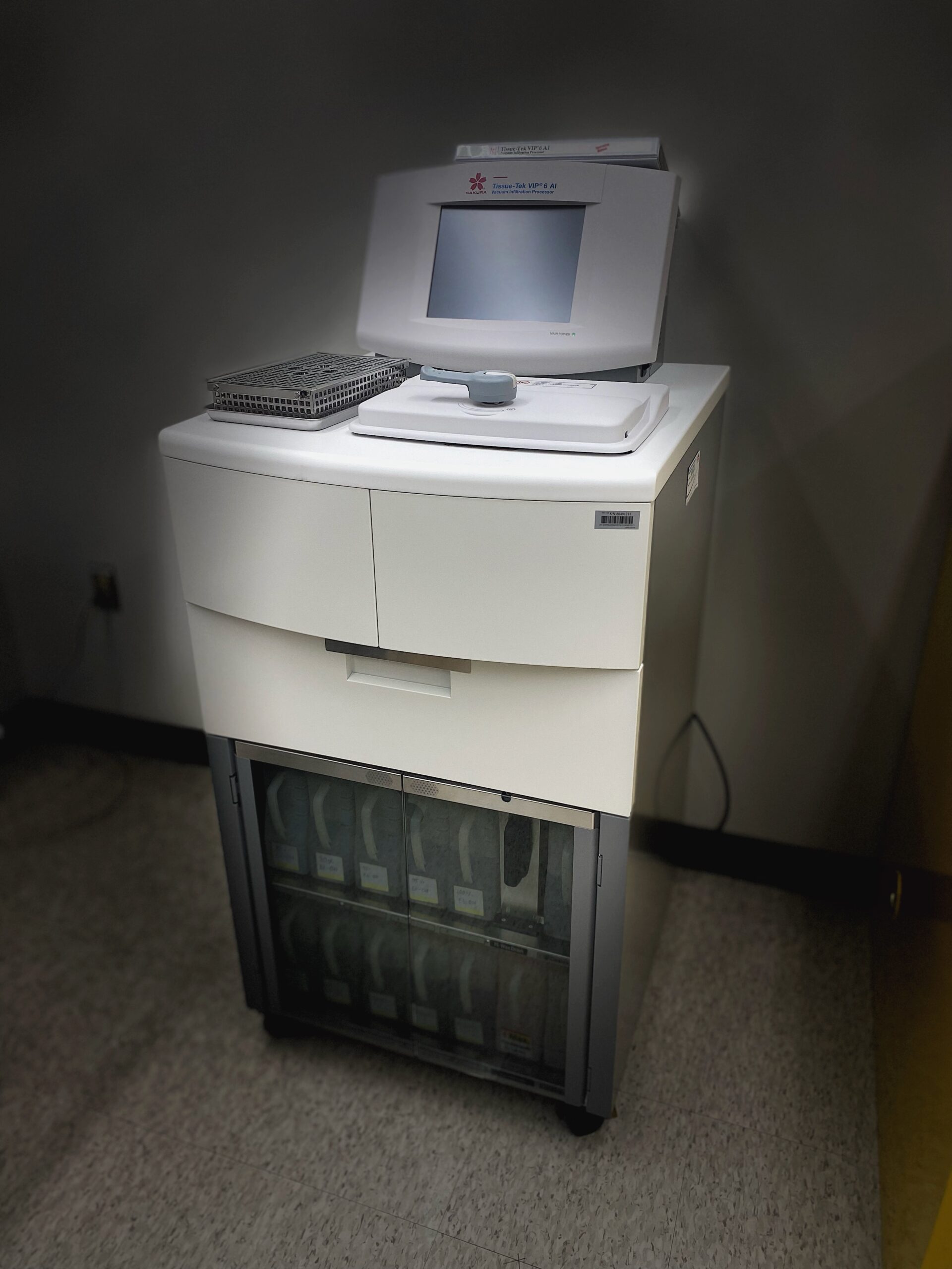
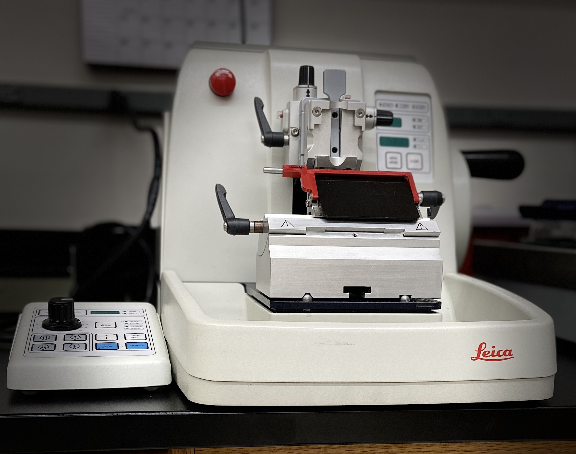
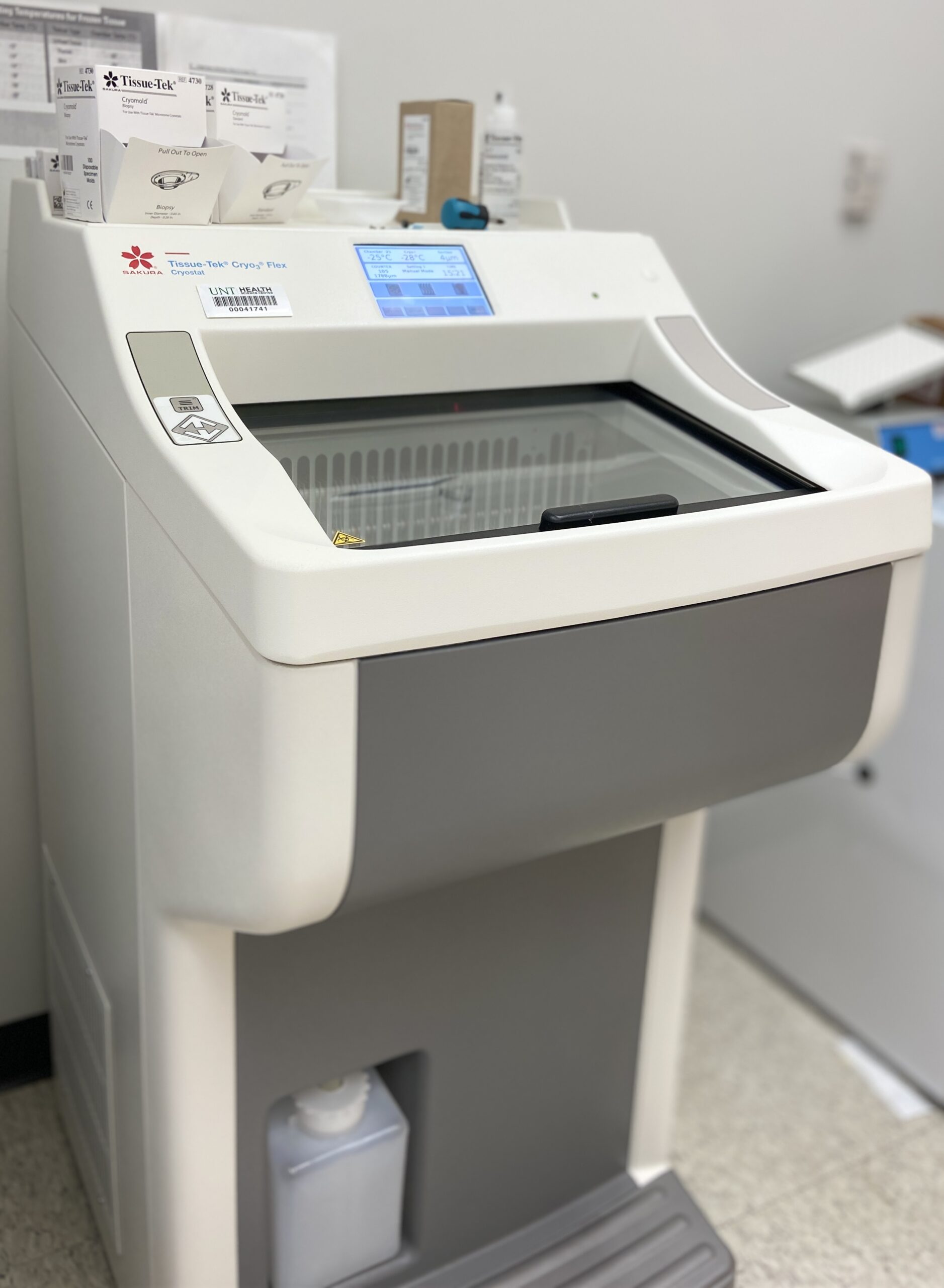
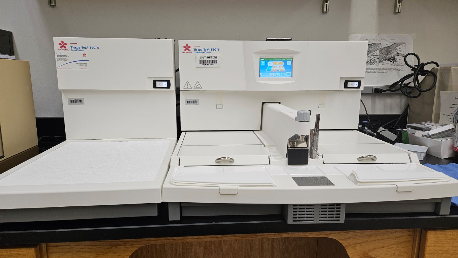
Full service Histology services from processing of tissue for Paraffin to Plastic embedding, sectioning and staining is provided by experienced histologist using up to date instrumentation like Tissue-Tek VIP AI Tissue Processor, Tissue-Tek Cryo3 Flex Cryostat, Leica Embedding station, Leica Microtomes, Leica UC7 Ultramicrotome. Training on equipment and techniques is provided by experienced histologist.
| Sharad Shrestha PhD | Director, Research Core | 817-735-0117 | Sharad.Shrestha@unthsc.edu |
| Kishor Kunwar MS | Manager, Microscopy Core | 817-735-0613 | Kishor.Kunwar@unthsc.edu |
| Hours | Location |
|
24/7 | Staffed Monday - Friday 8AM-5PM |
3500 Camp Bowie Blvd, RES 0121 & RES 0121A, EAD630 Fort Worth, TX 76107
|
| Name | Role | Phone | Location | |
|---|---|---|---|---|
| Kishor Kunwar, MSc |
Manager, Microscopy Core
|
817-735-0613
|
Kishor.Kunwar@unthsc.edu
|
RES 111
|
| Anne-Marie Brun |
Histologist
|
817-735-2045
|
Anne-Marie.Brun@unthsc.edu
|
EAD 630
|
| Tamara Mcinnis |
Histologist
|
tamara.mcinnis@unthsc.edu
|
EAD630
|
|
| Sharad Shrestha, PhD |
Research Core Labs Director
|
817-735-0117
|
Sharad.Shrestha@unthsc.edu
|